FEATURE: Scalp Inflammation: What We Test, What We Miss
Scalp discomfort is one of the most common complaints in clinic—itching, flaking, tightness, heat. And yet, it’s one of the least well understood.
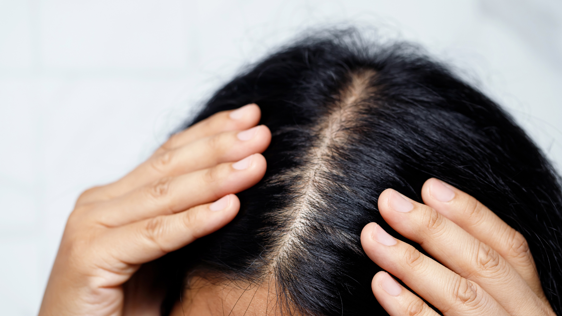
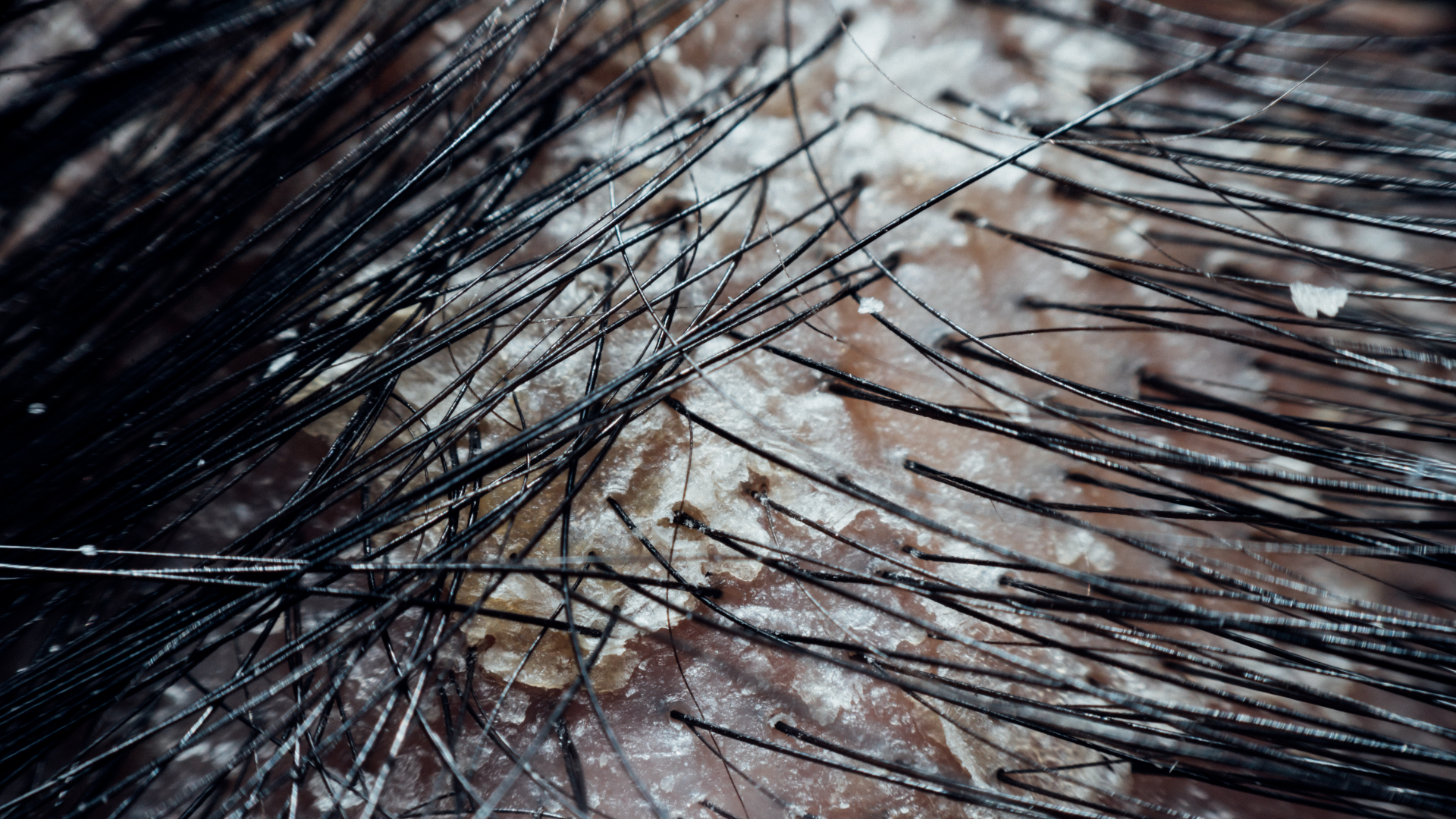
While acute or visibly inflamed conditions like psoriasis or seborrheic dermatitis tend to get clinical attention, many cases present without obvious signs—just a persistent irritation, or slow, generalised thinning with no scarring or redness.
These subtler cases are often brushed off, poorly investigated, or dismissed as cosmetic.

But there’s a growing body of evidence to suggest that chronic, low-grade inflammation—or “microinflammation”—may be silently disrupting follicular health in ways traditional diagnostics aren’t designed to detect.
When scalp inflammation is suspected, the standard pathway in most GP or dermatology settings usually involves four main approaches: scalp swabs, dermoscopy (trichoscopy), basic blood tests, and—less commonly—a biopsy.

Scalp Swab
A swab test involves collecting a sample from the scalp surface and sending it to a lab to identify bacterial or fungal overgrowth. It can detect infections like tinea capitis or Staphylococcus aureus, but it has limitations. Many common microbes (like Malassezia or Cutibacterium) aren’t reliably cultured this way. Inflammation caused by microbial imbalance—not overt infection—will often be missed entirely.
Trichoscopy
This is a visual inspection of the scalp using a dermatoscope. It helps assess scaling, vascular changes, follicular openings, or signs of scarring. While incredibly useful in cases like lichen planopilaris or frontal fibrosing alopecia, trichoscopy won’t pick up early-stage microinflammation unless the practitioner is looking specifically for subtle changes, like perifollicular erythema or loss of follicular uniformity.
Blood Tests
Standard lab panels may include full blood count (to screen for anaemia or infection), ferritin (iron storage), thyroid function, vitamin D, and B12. Occasionally, CRP or ESR will be checked to assess general inflammation. However, these tests are systemic—they do not tell you what’s happening locally at the scalp.
Biopsy
A punch biopsy can give a definitive histological diagnosis. It’s used to confirm inflammatory and scarring alopecias like lichen planopilaris or discoid lupus, and looks for signs like lymphocytic infiltration, perifollicular fibrosis, or immune cell clustering. But biopsies are rarely offered for mild, vague, or non-scarring cases. They’re invasive, delayed, and often reserved for complex or late-stage presentations.

The issue isn’t that these tools are useless—they’re just not built to detect subtle or chronic microinflammation. They’re designed to catch infection, autoimmunity, or obvious pathology. But what about the client who has slow-onset thinning, no redness, no scaling—but persistent scalp tension or discomfort?
In these cases, inflammation may be immune-mediated but non-destructive, affecting follicular signalling, vascular flow, and sebum quality without triggering a dramatic visual flare.
It’s inflammation that doesn’t scream—but still disrupts.
This is where functional testing and a more investigative approach become valuable:
Microbiome Analysis
Although not yet widely available for the scalp, shotgun or 16S sequencing is used in research settings to map the scalp’s microbial environment. These tests can identify shifts in bacterial balance—such as a loss of commensal Staphylococcus epidermidis or an overgrowth of Malassezia—that could be triggering localised inflammation without infection.
Functional Blood Markers
Nutrient panels assessing zinc, selenium, omega-3 fatty acids, homocysteine, and hs-CRP (high-sensitivity C-reactive protein) may reveal more subtle drivers of inflammation, oxidative stress, or poor barrier repair capacity—factors that directly impact scalp resilience.
Barrier Function Testing
While still emerging in clinical practice, some cosmetic science labs are using TEWL (transepidermal water loss)testing on the scalp to assess barrier integrity. A compromised barrier is a gateway for microbial imbalance, immune dysregulation, and chronic irritation.
Symptoms That May Point to Microbial Imbalance
☐ Itchy or tingling scalp with no visible cause
☐ Flaking that doesn’t respond to standard shampoos
☐ Burning or tightness after washing or product use
☐ Dry scalp with greasy roots
☐ Sensitivity to water, weather, or styling products
☐ Generalised thinning or shedding with “normal” test results
☐ Small bumps, biofilm-like residue, or recurrent irritation
☐ Scalp feels better after a break from products
☐Increase in hair porosity without chemical treatment
☐Brittle or dry hair texture
☐ Excessive scalp oiliness paired with dry ends
☐ Unusual or musty scalp odour linked to stress or diet change
☐ Delayed healing of scalp wounds or scratches
☐ Prolonged hair regrowth delay after shedding phase
☐ Flare-ups following sugar, alcohol, or processed food intake
☐ Itching or crawling sensations without a visible cause
Scalp inflammation isn’t always visible to the naked eye. And not all hair loss fits neatly into diagnostic boxes. If you’re experiencing discomfort, diffuse shedding, or texture changes—and your tests come back “normal”—it may be time to reframe the question. You’re not looking for a disease. You’re looking for a disruption.
Understanding and addressing microinflammation means going beyond symptoms and into the biology of the scalp as an ecosystem. And that starts with better testing, better tools—and better questions.


